
Diagnosis and Surgical Treatment
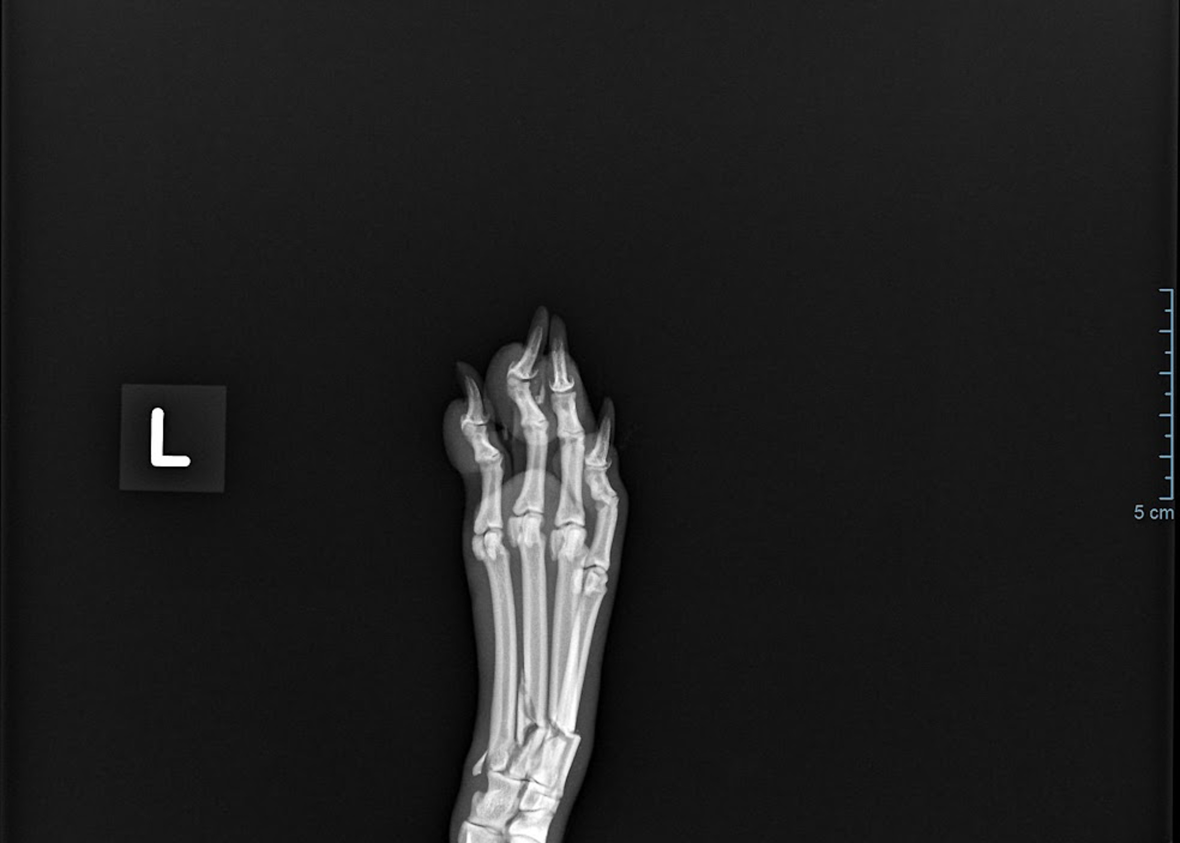
When is surgery recommended?
What signs might you notice?
Dogs and cats with metatarsal fractures may show:
• Sudden hindlimb lameness
• Reluctance or inability to bear weight
• Swelling of the paw
• Pain when the foot is handled
• Bruising
• Abnormal positioning of the toes
• Visible deformity of the paw
• Wounds following more severe trauma
The severity of lameness does not always indicate the complexity of the fracture, so radiographs are required to determine exactly which bones are affected.
How are metatarsal fractures diagnosed?
Diagnosis begins with examination of the injured limb, including assessment of pain, swelling, paw alignment, stability and the surrounding skin and soft tissues.
Radiographs are obtained in orthogonal projections to determine:
• Which metatarsal bones are fractured
• The location and configuration of each fracture
• The degree of displacement
• Alignment of the digits and paw
• Whether adjacent joints are involved
Where the injury follows major trauma, the patient is also assessed for other injuries before definitive fracture treatment is undertaken.
Do all metatarsal fractures require surgery?
No.
Stable fractures with little displacement may sometimes be treated using a splint or cast together with strict activity restriction.
Surgical stabilisation becomes more appropriate as displacement, instability and fracture complexity increase. Published evidence suggests external coaptation can produce good results in appropriately selected minimally displaced cases, while markedly displaced injuries are more likely to benefit from surgical restoration of alignment.
What are the metatarsal bones?
Dogs and cats normally have four principal weight-bearing metatarsal bones running parallel to one another within the hind paw.
Together they transfer load from the hock towards the digits during standing, walking and running. Although each bone is relatively small, the group functions as a unit and appropriate alignment is important for normal positioning of the toes and distribution of weight across the paw.
Trauma may fracture only one metatarsal or several bones simultaneously. When several adjacent bones are fractured, much of the normal internal support of the paw can be lost, creating considerable instability.
The Spider construct
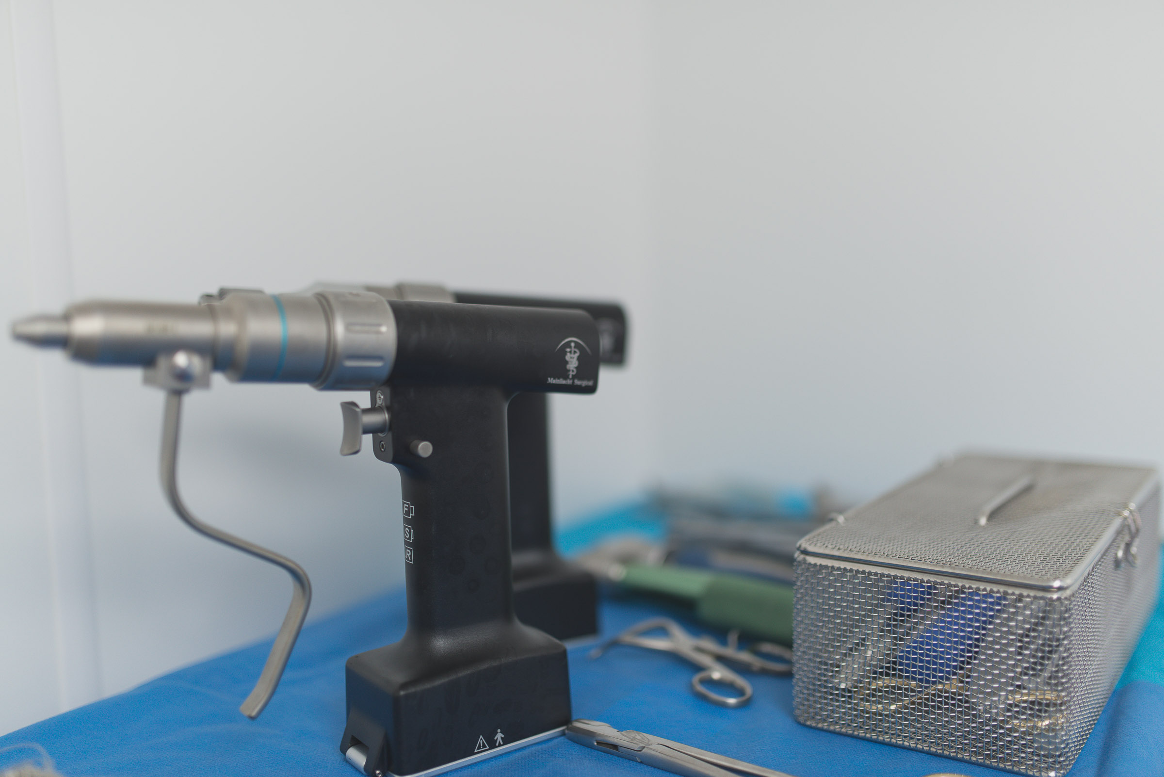
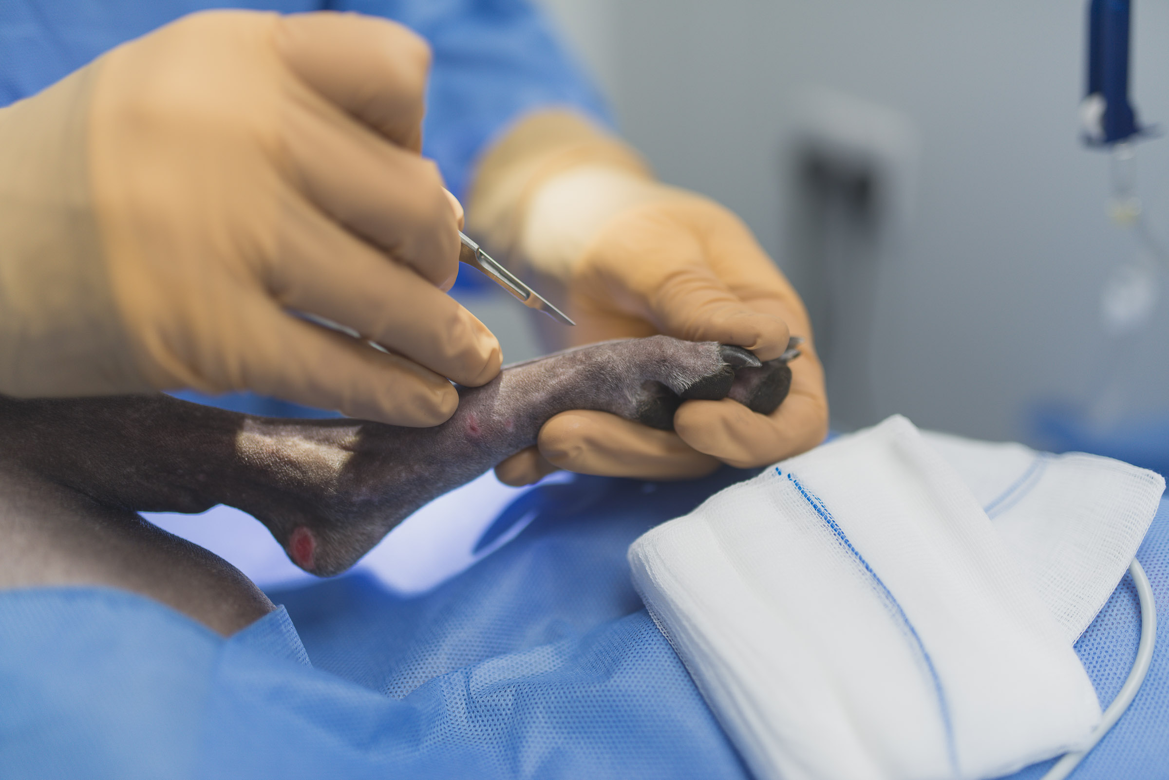
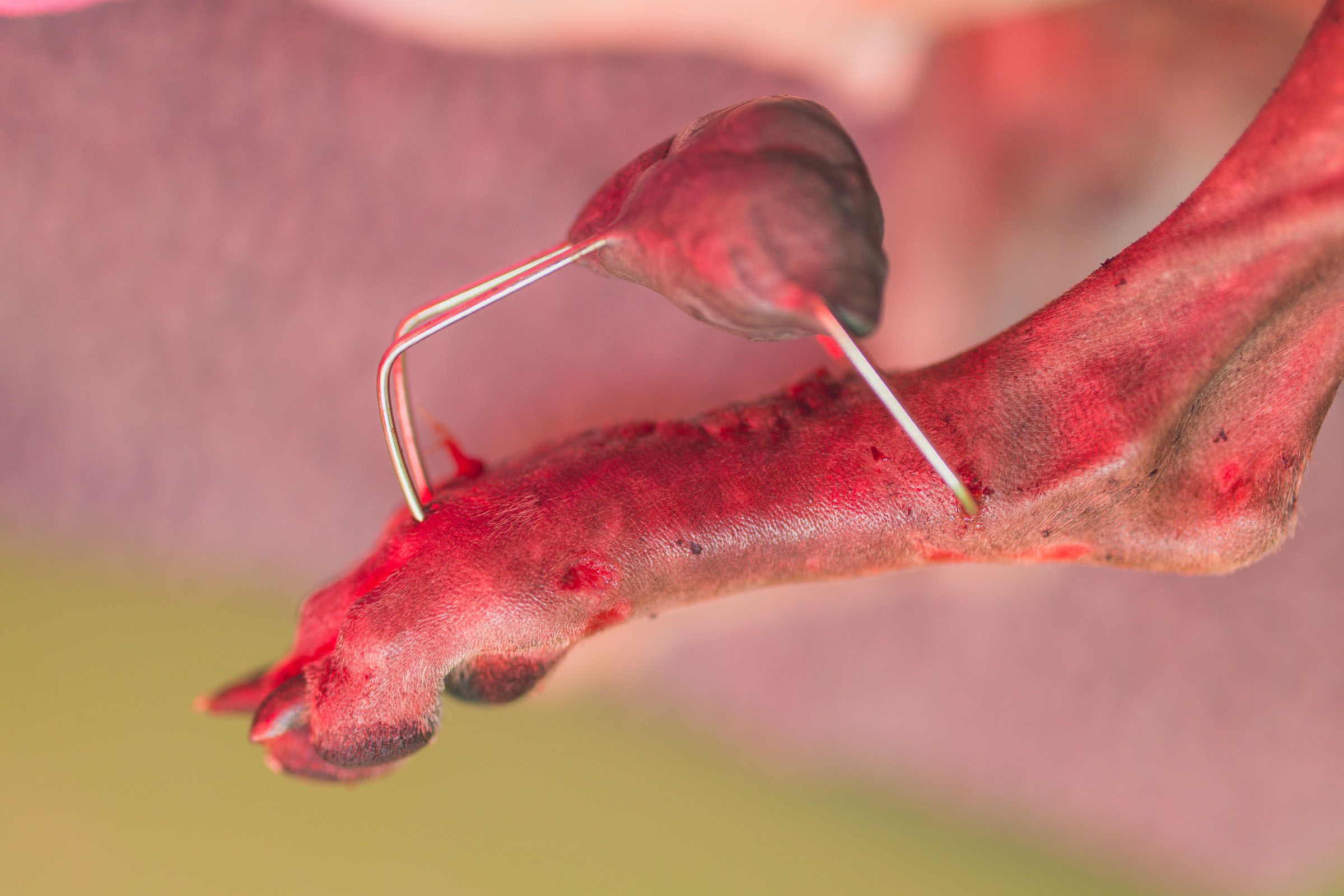
For selected complex metatarsal and metacarpal fractures, a combined intramedullary and external fixation technique sometimes referred to as a Spider construct can be used.
Small K-wires are placed to engage the fractured bones and are incorporated into an external supporting construct. This allows several small bones or short fracture fragments to be stabilised together while maintaining the overall alignment of the paw.
The technique can be particularly useful where conventional plate fixation of each individual metatarsal would be difficult or unnecessarily invasive.
A published series involving 12 dogs and 19 cats described combined intramedullary and external skeletal fixation for metacarpal and metatarsal fractures, with all frames removed within ten weeks. The authors considered the technique particularly useful for fractures close to the joints, although complications such as pin-tract discharge, swelling and frame irritation can occur.
What happens after surgery?
After fixation has been completed, the stability and alignment of the paw are assessed and postoperative radiographs are obtained.
The patient is monitored during recovery and appropriate pain relief is provided.
Depending on the fracture configuration and fixation technique, additional padded support may be used during the early postoperative period.
Patients are normally discharged with detailed instructions covering activity restriction, medication, wound or pin-site monitoring and scheduled rechecks.
Post-operative care
Recovery at home is an important part of successful fracture treatment.
During the early healing period:
• Activity should be strictly restricted
• Exercise should be controlled and kept short
• Running and jumping should be prevented
• Dressings or external fixation components must be kept clean and dry
• The paw should be checked regularly for swelling or discomfort
• All prescribed medications should be given as directed
Patients are re-examined during recovery, and repeat radiographs are taken to assess fracture healing.
Where an external construct has been used, removal is planned once sufficient healing has occurred rather than according to a fixed date.
Risks and complications
All fracture repairs carry some risk.
Potential complications following metatarsal fracture surgery include:
• Swelling and bruising
• Wound complications
• Infection
• Pin-tract irritation or discharge
• Implant loosening or movement
• Loss of fracture alignment
• Delayed bone healing
• Non-union
• Bridging of adjacent metatarsal bones during healing
• Irritation caused by an external frame
• Need for additional surgery
Complications involving pin tracts and frame irritation are recognised with external fixation techniques, although published case series have also reported good long-term limb function following successful healing
Recovery and expected outcome
Recovery depends on the severity of the original injury, number of bones affected, patient age and fixation technique.
0–2 weeks
Strict activity restriction while early soft tissue and bone healing begins. Wounds, dressings and any external fixation components are monitored closely.
2–6 weeks
Controlled activity remains important. Clinical rechecks are performed and limb use should gradually improve as healing progresses.
6–10 weeks
Repeat radiographs are commonly used to assess progression of bone healing. External fixation may be removed when adequate healing has occurred.
Long term
Exercise is gradually increased once fracture healing has been confirmed. The aim is comfortable weight bearing and return to normal or near-normal function.
Long-term prognosis for metacarpal and metatarsal fractures is generally favourable, although greater displacement and instability are associated with increased complication risk. In one retrospective series of 100 dogs, persistent clinical lameness was uncommon at long-term reassessment. PubMed
Frequently Asked Questions